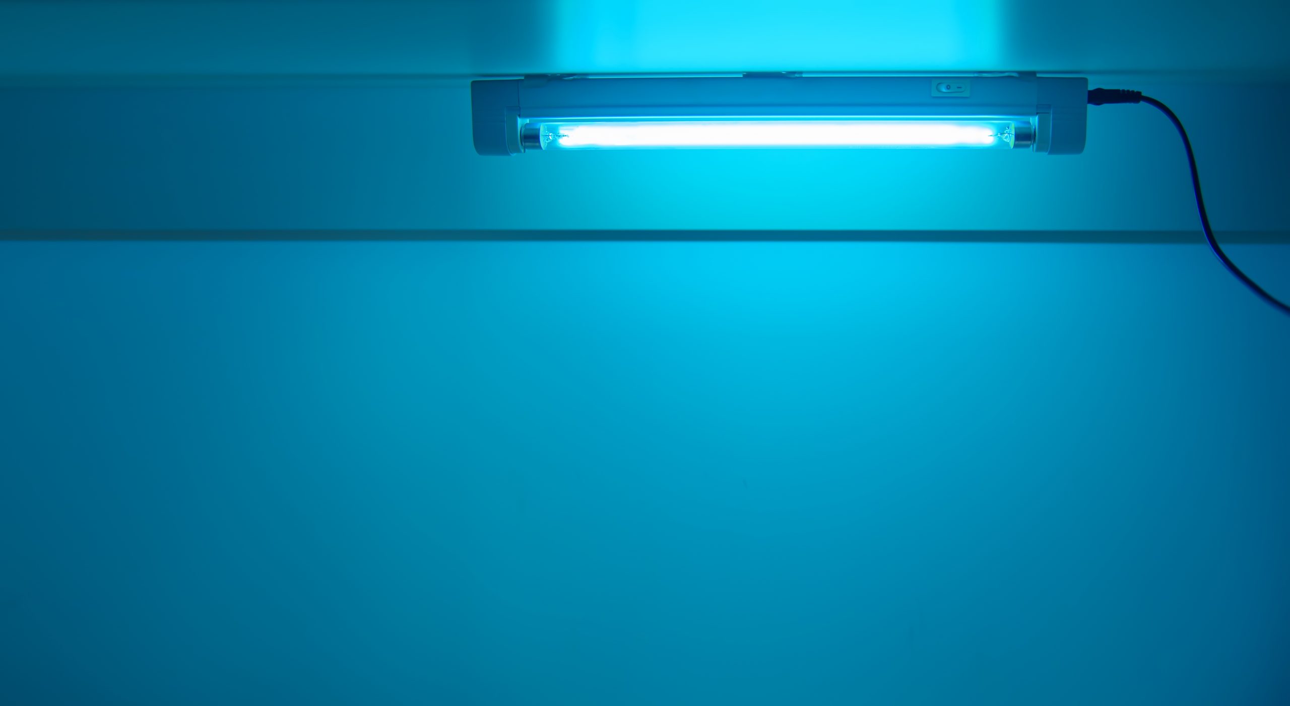
Ultraviolet (UV) irradiation has been known for decades as a terminal disinfection method, widely used in a variety of applications including healthcare, agriculture, water and food treatment, as well as in airlines and many other settings. UV radiation covers the wavelength range from 100 to 400 nanometers (nm). This is further divided into four distinct spectral areas including: UV-A (315–400 nm), UV-B (280–315 nm), UV-C (200–280 nm) and vacuum UV (100–200 nm). Among these wavelength ranges, UV-C has been evidently shown to have the highest efficacy against microorganisms. UV-C wavelength, 250–270nm, is strongly and primarily absorbed by the nucleic acids of microbial cells leading to DNA disruption and cell death. UV-C are known to be a safer choice compared to UV-A and UV-B light that were shown to be carcinogenic. This is because UV-C radiation is absorbed by the outer dead layer of human skin, while UV-B and UV-A radiation penetrate deeper. Different UV sources have been utilized such as low and medium pressure mercury UV lamps, UV light-emitting diodes (UV-LEDs), and far-UVC (207–222 nm) radiating excimer and micro-plasma lamps as well as broad spectrum technologies such as pulsed xenon that applies the whole UV spectrum (UV-A, UV-B and UV-C).
The microbiocidal mechanism of UV-C is to cause damage to their RNA and DNA, which often leads to the formation of dimers between pyrimidine residues in the nucleic acid strands. The consequence of this modification is that the production of cyclobutane pyrimidine dimers (CPDs) causes deformation of the DNA or RNA molecule, which might cause defects in cell replication leading to cell death afterwards. UV light causes physical electron movements and destroys DNA bonds. UV light induces the formation of photoproducts due to the direct absorption of photons by pyrimidine and purine nucleic acid bases. Photoproducts lead to structural distortion in DNA and interrupt RNA transcription and DNA replication, finally causing cell mutagenesis or death. The major photoproducts caused by UV are cyclobutane pyrimidine dimers (CPDs) and pyrimidine 6-4 pyrimidone photoproducts (6-4 PPs).

Before proceeding to the efficacy of UV-C against a wide range of microorganisms, we must understand the kinetics and parameters of UV-C irradiation. FUV decontamination is an energy-based process, meaning that the microbial inactivation rate is determined by the delivered UV dose via the decontaminating unit. Mcdevitt and collegues (2012) stated that microorganism susceptibility to UV-C light is traditionally thought to follow first-order kinetics according to the equation FR= CUV/CNo UV= e-ZD, where FR is the fraction remaining, CUV is microorganism concentration with UV exposure, CNo UV is microorganism concentration without UV exposure, Z is the susceptibility parameter expressed in m²/J, and D is the dose of UV-C in J/m² or mJ/cm² (milli second joule per square centimeter). In other words, the UV dose is the product of UV intensity (expressed as energy per unit surface area) and residence time. These susceptibility parameters, or Z values, do not appear to be static but vary with variable conditions, such as relative humidity (RH), microbial sensitivity and target medium characteristics.
Multiple trials have demonstrated the effectiveness of ultraviolet (UV-C) light devices in reducing the environmental bioburden of pathogenic organisms such as methicillin-resistant Staphylococcus aureus (MRSA), vancomycin-resistant Enterococcus spp (VRE), Clostridium difficile, Acinetobacter spp., H1N1 Influenza virus, noroviruses and coronaviruses. However, microbial sensitivity to UV radiation varies widely. For instance, the rotavirus requires about 25 mJ/cm² of 254 nm UVC radiation from a mercury discharge lamp for a 3-log reduction, but for adenovirus (Type 40), the value is approximately 6 times higher (140 mJ/cm²).

To further support these claims, a study by Kim and colleagues(2015), reported that decontamination efficacy was more dependant to dose level than to the wavelength measurements. UV-LEDs achieved 5-log reductions of E. coli O157:H7 after 0.5 mJ/cm² and S. typhimurium after 0.7 mJ/cm² , and in the case of L. monocytogenes they achieved 5-log reductions after 0.7 mJ/cm² only at 266 and 270 nm. Higher dose level was needed for L. monocytogenes (gram-positive bacteria) to achieve the same reduction of E. coli O157:H7 or S. Typhimurium (gram-negative bacteria). Gram-positive bacteria are generally more resistant to UV light than are Gram-negative bacteria. This was demonstrated by the study of Beauchamp and Lacroix (2012), who reported that L. monocytogenes produced 35% fewer CPDs and 10% fewer 6-4 PPs than E. coli during a UV lamp irradiation dose of >3 mJ/cm² . This low production of UV photoproducts indicates a greater resistance for Gram-positive bacteria.
A recently published study in the American Journal of Infection and Control, determined the susceptibility of SARS-COV-2 to UV-A or UV-C irradiation. Viral stock at a concentration of 5 x 10⁶ TCID50/mL was irradiated with UV light for up to 30 minutes. UV exposure was performed by separate or combined irradiation with UV-C (254 nm) and/ or UV-A (365 nm) of 600 µL virus stock in 24-well plates. The UV light source was placed at 3 cm above the bottom of the plate. The emitted light intensity was UV-C (254 nm) = 1940 mW/cm² and UV-A (365 nm) = 540 mW/cm² at 3 cm distance, as measured by radiometric analysis. This corresponds to an applied light dose of 1.94 mJ/ cm² per second for UV-C and 0.54 mJ/cm² per second for UV-A.
SARS-CoV-2 showed high susceptibility to UV irradiation. UV-A exposure alone was less effective on virus inactivation. After 9 minutes of irradiation to an emitted dose of 292 mJ/cm², 1 log reduction of the viral load was observed. In contrast, complete virus inactivation was achieved after 9 minutes exposure to UV-C to an emitted dose of 1048 mJ/cm². These data confirm former findings that UV-C is more effective in inactivating viruses and highlights UV-C irradiation as an effective method for the inactivation of SARS-CoV-2.

It is necessary to determine Z values in a defined and controlled experimental system to predict UV-C effectiveness, determine the amount of UV-C energy required for intervention, and develop models of UV-C systems. For example, the influenza Z values were much higher (i.e., influenza is more susceptible to UV-C) than those reported for hardy spores, such as those from Bacillus anthracis. Kowalski and colleagues (2000) were able to calculate a Z value of 0.15 m² /J. The differences in Z values between both data and previously reported data, although modest, may correspond to large differences in virus survival.
In contrast, Mcdevitt and colleagues (2012) showed that a dose of 6 J/m² and a Z value of 0.15 m²/J would result in a 60% reduction, while a Z value of 0.25 would result in a 77% reduction for influenzas virus. The Z values for influenza virus were statistically different as a function of RH. These differences showing that influenza aerosols are less susceptible to UV at higher RH than at lower RH have been noted in previous UV susceptibility studies for other organisms. In climate-controlled, indoor environments, high RH values near 75% may be unusual, but in areas without environmental controls UV effectiveness may decrease in the presence of elevated RH.
Based on the findings from the literature review, some parameters are critical and should be monitored to ensure efficacy. These parameters include the RH, microbial sensitivity, irradiance exposure and distance. Which all contribute to successful cycle developments and efficacy. A good system then should have a technology that is able to monitor all these parameters in order to ensure efficacy of microbial inactivation has been achieved with minimum exposure of hazards as well as performing automated adjustments when needed.

UV-C has been praised numerously as an effective and safe decontamination method in treating a wide variety of contaminated settings and objects. It has shown effectiveness against a wide variety of microorganisms including the novel coronavirus which was reported to be one of the most susceptible viruses to UV light. The differences in microbial characteristics and environmental conditions highly determined the appropriate Z values that are required to achieve inactivation. Air aerosols carrying Influenza virus were shown to require higher dosages of UV-C to be inactivated in comparison with surfaces. This is because RH plays a critical role in protecting the pathogens from UV light. The higher the RH the lower the Z value which is in turn dependant to each type of microorganism. Therefore, it is pivotal for the decontamination technology to determine RH levels of the environment and the Z values of a particular pathogen prior to testing the UV-C efficacy of a specific dose.